
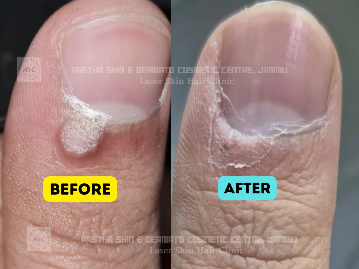
Dermatologist assessment before removing any skin growth
Consult Dr. Cheena Langer, MBBS, MD Dermatology, Consultant Dermatologist, for diagnosis and removal of suitable warts, moles and skin tags at Aastha Skin & Dermato-Cosmetic Centre in Jammu.
Many skin growths look similar. A suspected skin tag may be a wart, mole, seborrhoeic keratosis or another lesion. Examination should therefore come before cautery, freezing, cutting or any home-removal attempt.
Wart, mole and skin-tag removal is available at both the Karan Nagar and Paloura branches.
What is a wart?
A wart is a usually non-cancerous skin growth caused by infection with certain types of human papillomavirus, or HPV. Warts can develop on the hands, feet, face, around nails and other body areas.
Warts may appear:
- Rough and raised
- Flat and smooth
- Thread-like
- Thickened on the sole
- In clusters
- With small dark dots
- Around fingers or nails
Warts can spread to other parts of the body or to another person through direct contact and contaminated personal items. Picking, shaving over or cutting a wart can contribute to spread.
How are ordinary skin warts treated?
The choice depends on the wart type, location, number, patient’s age, medical history and previous treatment.
Options a dermatologist may consider include:
- Salicylic-acid treatment
- Cryotherapy or freezing
- Electrosurgery
- Curettage
- Excision
- Selected prescription medicines
- Immunotherapy for resistant warts
- Laser treatment for selected stubborn cases
Cryotherapy commonly causes a blister or crust, and several sessions may be needed. Electrosurgery and curettage destroy and remove the wart after the skin is numbed. Excision removes it immediately but can leave a scar.
No method can promise that a wart will never return because treatment removes the visible growth but the virus may persist in surrounding skin.
When should a wart be examined by a dermatologist?
Book an assessment when:
- The diagnosis is uncertain
- The wart is on the face
- It is painful, itchy or bleeding
- It is changing rapidly
- Many warts are present
- Treatment has repeatedly failed
- The patient has diabetes
- Immunity is reduced
- The lesion looks like an open sore
- It develops around or beneath a nail
A rapidly growing, bleeding or treatment-resistant lesion may require biopsy to confirm that it is a wart rather than another skin condition.
What is a mole?
A mole, medically called a naevus, is a growth formed by pigment-producing cells.
Moles may be:
- Brown
- Black
- Tan
- Pink
- Skin-coloured
- Flat
- Raised
- Smooth
- Hair-bearing
Most moles are harmless and do not require removal. A dermatologist may remove a mole when it is repeatedly irritated, cosmetically concerning or suspicious for skin cancer.
Types, options and detailed guidance
Common warts
Common warts usually appear as rough bumps on the hands, fingers, knees or elbows.
Plantar warts
Plantar warts develop on the soles. Pressure from walking may push them inward and create pain or a callus-like surface.
Flat warts
Flat warts are smaller and smoother. They may occur in larger numbers on the face, neck, beard area or legs.
Filiform warts
Filiform warts have a narrow, thread-like appearance and commonly affect the face or neck.
Periungual warts
These occur around or beneath the nails and may disturb nail growth.
Diagnosis should be confirmed because corns, calluses, skin cancers and other growths can sometimes resemble warts.
Are genital warts treated differently?
Yes. Genital and perianal warts require a separate medical assessment.
Do not apply ordinary over-the-counter hand or foot wart medicines to genital skin. Treatment may involve prescription medicine, cryotherapy, electrocautery, excision or another procedure selected according to the number and location of the lesions.
Patients should also receive confidential counselling about HPV transmission, sexual health and relevant screening. Removing visible genital warts does not necessarily eliminate HPV, so recurrence and transmission can still occur.
When should a mole be checked?
Arrange a dermatology examination when a mole:
- Changes in size
- Changes in shape
- Changes in colour
- Becomes itchy
- Starts bleeding
- Develops crusting
- Becomes painful
- Looks different from the person’s other moles
- Appears newly in adulthood
- Grows rapidly
A changing, itching or bleeding mole can be a warning sign of melanoma and should not be removed cosmetically without proper assessment.
What are the ABCDE warning signs?
The ABCDE guide can help identify a mole that needs examination:
- A – Asymmetry: one half differs from the other
- B – Border: irregular, blurred or poorly defined edge
- C – Colour: several colours or uneven pigmentation
- D – Diameter: often larger than 6 mm, although melanoma can be smaller
- E – Evolving: changing in size, shape, colour or symptoms
A lesion that looks unlike the patient’s other moles, the “ugly duckling”, also deserves assessment.
The ABCDE guide does not replace examination. Some melanomas do not follow every rule.
How are moles removed?
The removal method depends on:
- Clinical appearance
- Size
- Depth
- Body location
- Cosmetic expectations
- Whether laboratory examination is required
- Tendency to form scars or keloids
Shave removal
A raised mole may be carefully shaved level with the surrounding skin. The sample may be sent for histopathology when appropriate.
Surgical excision
The entire lesion is cut out after local anaesthesia. Stitches may be required.
Diagnostic biopsy
When the mole is suspicious, the priority is obtaining an adequate sample for diagnosis rather than choosing the quickest cosmetic technique.
The AAD notes that surgical excision and surgical shaving are commonly used for mole removal.
Will a removed mole be sent for testing?
It may be.
Histopathology means examining removed tissue under a microscope. It may be recommended when:
- The mole is changing
- The diagnosis is uncertain
- The colour or border is irregular
- The lesion has bled
- It looks different from other moles
- The doctor needs to exclude skin cancer
- The method of removal produces a suitable specimen
Suspicious growths should not simply be burnt or destroyed without considering whether tissue is needed for diagnosis. A dermatoscope and skin biopsy may be used when melanoma is suspected.
Skin-biopsy services are available at both Aastha Skin Centre locations.
Electrosurgery or electrodesiccation
Controlled electrical energy destroys the growth. A small crust commonly forms and heals gradually.
Snip removal
After cleaning and numbing the area where required, the tag is removed using sterile surgical scissors or a blade.
Cryotherapy
The lesion is frozen using a very cold substance. It later crusts or falls away.
The appropriate technique depends on the location, stalk width, bleeding risk and possibility that the lesion is not a skin tag.
Additional care and practical advice
Diagnosis before destruction
The lesion is examined before it is frozen, cauterised, shaved or excised.
Dermatologist-led care
Assessment is provided under Dr. Cheena Langer, MBBS, MD Dermatology, Consultant Dermatologist.
Multiple lesion types evaluated
The clinic assesses warts, moles, skin tags, DPN, seborrhoeic keratoses and other skin growths.
Biopsy when indicated
Suspicious or uncertain lesions can be considered for tissue examination rather than treated only as a cosmetic concern.
Two Jammu locations
Wart, mole and skin-tag removal is confirmed at both Karan Nagar and Paloura.
Realistic counselling
Patients receive information about healing, recurrence, pigmentation and possible scarring before treatment.
How care is planned
- Assessment comes before treatment selection.
- Options, limitations, aftercare and expected timelines are explained.
- The plan is adapted to diagnosis, skin type and medical history.
Choose a clinic in Jammu
Karan Nagar
Lane 2, Karan Nagar, near Amphalla Chowk, Jammu – 180005Doctor consultation:
Mon–Sat 11:00 AM–4:00 PM
Sun 11:00 AM–3:00 PM
Paloura Chowk
Top Paloura, opposite Government Senior Secondary School, Jammu – 181121Doctor consultation:
Mon–Sat 6:00 PM–8:00 PM
Sun 10:30 AM–12:00 PM
Frequently asked questions
Can a mole grow back after removal?
A mole can recur when some mole cells remain in the skin, particularly after a superficial shave procedure.
Recurrence does not automatically mean cancer, but the regrowing area should be examined, especially if it develops irregular colour, bleeding, rapid growth or another change.
Complete excision may reduce recurrence but usually creates a longer scar and may require stitches. The procedure should therefore be selected according to diagnosis, depth and cosmetic priorities.
What is a skin tag?
A skin tag is a soft, usually harmless growth that commonly hangs from the skin on a small stalk.
Skin tags frequently occur on:
- Neck
- Underarms
- Eyelids
- Groin
- Beneath the breasts
- Other friction-prone folds
They may be skin-coloured or darker and can become irritated by jewellery, clothing or shaving.
A dermatologist may remove a skin tag using cryosurgery, electrodesiccation or sterile snip removal, depending on its size and location.
Do skin tags require treatment?
Most skin tags do not require medical treatment.
Removal may be considered when a tag:
- Rubs against clothing
- Gets caught in jewellery
- Becomes repeatedly irritated
- Bleeds after friction
- Interferes with shaving
- Causes cosmetic concern
- Has an uncertain diagnosis
Sudden development of numerous growths or an unusual-looking lesion should be assessed rather than automatically assumed to be an ordinary skin tag.
How are skin tags removed?
Depending on the lesion, a dermatologist may use:
Electrosurgery or electrodesiccation
Controlled electrical energy destroys the growth. A small crust commonly forms and heals gradually.
Snip removal
After cleaning and numbing the area where required, the tag is removed using sterile surgical scissors or a blade.
Cryotherapy
The lesion is frozen using a very cold substance. It later crusts or falls away.
The appropriate technique depends on the location, stalk width, bleeding risk and possibility that the lesion is not a skin tag.
Why should skin tags not be removed at home?
Patients should avoid cutting, tying, burning or applying unregulated acids to a presumed skin tag.
Home removal can cause:
- Bleeding
- Infection
- Chemical burns
- Pigmentation
- Scarring
- Injury to surrounding skin
- Incomplete removal
- Delay in diagnosing another skin growth
A dark or changing lesion may not be a skin tag. Destroying it at home can eliminate tissue needed for diagnosis.
What are DPN and seborrhoeic keratoses?
Seborrhoeic keratoses are common, usually harmless growths that may look waxy, rough or “stuck on” to the skin.
Dermatosis papulosa nigra, or DPN, describes multiple small, dark, raised facial or neck growths that commonly occur in deeper skin tones. Diagnosis should be confirmed before cosmetic removal.
Seborrhoeic keratoses usually need no treatment, but they may be removed if irritated, cosmetically concerning or difficult to distinguish from another lesion. Options may include cryotherapy, electrosurgery, curettage or shave removal.
DPN and seborrhoeic keratosis removal are confirmed as available at both Aastha Skin Centre branches.
How does Dr. Cheena Langer assess a skin growth?
A consultation may include:
- Reviewing when the growth first appeared
- Assessing whether it has changed
- Asking about bleeding, itching or pain
- Examining its shape, colour and surface
- Checking whether similar growths are present elsewhere
- Reviewing previous removal attempts
- Assessing scar or keloid tendency
- Considering dermoscopy
- Deciding whether biopsy is needed
- Discussing the appropriate removal method
A growth may be observed rather than removed when treatment is unnecessary or the cosmetic risks outweigh the likely benefit.
Get a clear plan for wart, mole & skin tag removal
Request a consultation with Dr. Cheena Langer at Karan Nagar or Paloura Chowk.
Medical disclaimer: Results may vary from person to person. This content is for informational purposes only and does not substitute a consultation with a qualified medical professional. Dr. Cheena Langer, MD is a registered medical practitioner.